Home » Uncategories » Upper Back Anatomy / How To Draw The Upper Back Anatomy And Motion Proko - The rib cage also anchors the bones of the head, neck, shoulders, and arms to the trunk of the body.
Upper Back Anatomy / How To Draw The Upper Back Anatomy And Motion Proko - The rib cage also anchors the bones of the head, neck, shoulders, and arms to the trunk of the body.
Upper Back Anatomy / How To Draw The Upper Back Anatomy And Motion Proko - The rib cage also anchors the bones of the head, neck, shoulders, and arms to the trunk of the body.. Toward the bottom, toward the belly superior: Upper back pain is most commonly caused by muscle irritation or tension, also called myofascial pain. After, behind, following, toward the rear distal: It runs from the neck to the upper back. The back is the body region between the neck and the gluteal regions.
See upper back stock video clips. It is like that for several reasons, all of which you can understand by looking at the anatomy of the thoracic spine. The shoulder joint is formed where the humerus (upper arm bone) fits into the scapula (shoulder blade), like a ball and socket. The main superficial muscles of the back are the following: Some of the exercises which an individual can do to relax or loosen the upper neck and back muscles are:
Diagram Back Muscles Human Anatomy Diagram Anatomia Muscular Anatomia Do Corpo Humano Corpo Humano from i.pinimg.com After, behind, following, toward the rear distal: Other important bones in the shoulder include: The rib cage also anchors the bones of the head, neck, shoulders, and arms to the trunk of the body. The cervical spine supports the weight and movement of your head and protects the nerves exiting your brain. Upper right back pain under shoulder blade. Away from, farther from the origin proximal: The cervical spine has 7 stacked bones called vertebrae, labeled c1 through c7. Spinal cord anatomy model 13 photos of the spinal cord anatomy model activate javascript how important are the spinal cord and the spinal nerves, labeled spinal cord model, nerves of spinal cord, spinal cord cross section, spinal cord quiz, spinal nerve model, spinal nerve numbering, what are the 31 spinal nerves, human anatomy, how important …
Muscle relaxation anatomy 12 photos of the muscle relaxation anatomy muscle relaxation anatomy, steps of muscle relaxation anatomy, human muscles, muscle relaxation anatomy, steps of muscle relaxation anatomy
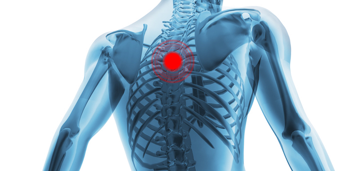
Upper right back pain under shoulder blade. There is a set of muscles in the upper back (called the thoracic area) called the spinalis thoracis. The rib cage also anchors the bones of the head, neck, shoulders, and arms to the trunk of the body. Spinal cord anatomy model 13 photos of the spinal cord anatomy model activate javascript how important are the spinal cord and the spinal nerves, labeled spinal cord model, nerves of spinal cord, spinal cord cross section, spinal cord quiz, spinal nerve model, spinal nerve numbering, what are the 31 spinal nerves, human anatomy, how important … Away from, farther from the origin proximal: Near, closer to the origin dorsal: The iliocostalis muscles are furthest from the spine. Before giving our recommendations for upper back exercises, it's important to first go over the anatomy of the back musculature. Other important bones in the shoulder include: In front of, front posterior: Powerful muscles that move the head and arms attach to these bones as well. Muscle strain, sprains, and spasms can affect the rhomboid muscles, which are located in the middle of the shoulder blades.this pain is mostly felt in. The complexity of this region means that dysfunction can occur either due to injury or progressive pain and degeneration.
The muscles of the chest and upper back occupy the thoracic region of the body inferior to the neck and superior to the abdominal region and include the muscles of the shoulders. Other important bones in the shoulder include: The cervical spine has 7 stacked bones called vertebrae, labeled c1 through c7. Powerful muscles that move the head and arms attach to these bones as well. Upper right back pain under shoulder blade.
Blog Causes Of Upper Back Pain from www.precisionpaincarerehab.com It consists of seven vertebrae. These include the cervical vertebrae in the neck, the thoracic vertebrae of the ribcage in the upper and middle back, the lumbar vertebrae in the lower back, and the vertebrae that are part of the pelvis. Other important bones in the shoulder include: This is a tutorial to quickly s. Back muscles anatomy here include the trapezius, latissimus dorsi, rhomboid and levator scapulae. Upper back pain is most commonly caused by muscle irritation or tension, also called myofascial pain. In the upper back region, the trapezius, rhomboid major, and levator scapulae muscles anchor the scapula and clavicle to the spines of several vertebrae and the occipital bone of the skull. Muscle strain, sprains, and spasms can affect the rhomboid muscles, which are located in the middle of the shoulder blades.this pain is mostly felt in.
This is a tutorial to quickly s.
The iliocostalis muscles are furthest from the spine. See upper back stock video clips. Powerful muscles that move the head and arms attach to these bones as well. The cervical spine is the top part of the spine. After, behind, following, toward the rear distal: It consists of seven vertebrae. The nervous system of the thorax is a vital part of the nervous system as a whole, as it includes the spinal cord, peripheral nerves, and autonomic ganglia that communicate with and control many vital organs. All these muscles are therefore associated with movements of the upper limb. The cause may be poor posture (such as forward head posture) or any type of irritation of the large back and shoulder muscles, including muscle strain or spasms. Before giving our recommendations for upper back exercises, it's important to first go over the anatomy of the back musculature. The main superficial muscles of the back are the following: Looking for a solution to your back pain problem? The bones of the chest and upper back combine to form the strong, protective rib cage around the vital thoracic organs such as the heart and lungs.
Upper right back pain under shoulder blade. As viewed from the side, the cervical spine forms a lordotic curve by gently curving toward the front of the body and then back. In front of, front posterior: The upper back muscles of the rhomboids and the trapezius are responsible for many of the movements of the scapula which in turn plays a huge role in the stability and mobility of the shoulder. These include the cervical vertebrae in the neck, the thoracic vertebrae of the ribcage in the upper and middle back, the lumbar vertebrae in the lower back, and the vertebrae that are part of the pelvis.
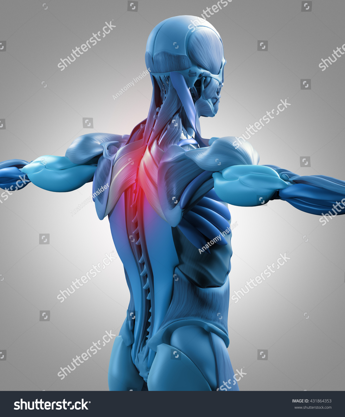
Human Anatomy Muscle Groups Torso Back Stock Illustration 431864353 from image.shutterstock.com The complexity of this region means that dysfunction can occur either due to injury or progressive pain and degeneration. The cervical spine supports the weight and movement of your head and protects the nerves exiting your brain. Looking for a solution to your back pain problem? It comprises the vertebral column (spine) and two compartments of back muscles; Some of the exercises which an individual can do to relax or loosen the upper neck and back muscles are: Back muscles anatomy here include the trapezius, latissimus dorsi, rhomboid and levator scapulae. Extending from the base of the skull into the pelvis, it consists of 33 stacked bones known as vertebrae. Muscle relaxation anatomy 12 photos of the muscle relaxation anatomy muscle relaxation anatomy, steps of muscle relaxation anatomy, human muscles, muscle relaxation anatomy, steps of muscle relaxation anatomy
As viewed from the side, the cervical spine forms a lordotic curve by gently curving toward the front of the body and then back.
Toward the bottom, toward the belly superior: The upper back muscles of the rhomboids and the trapezius are responsible for many of the movements of the scapula which in turn plays a huge role in the stability and mobility of the shoulder. Near, closer to the origin dorsal: The cervical spine is the top part of the spine. Extending from the base of the skull into the pelvis, it consists of 33 stacked bones known as vertebrae. These include the cervical vertebrae in the neck, the thoracic vertebrae of the ribcage in the upper and middle back, the lumbar vertebrae in the lower back, and the vertebrae that are part of the pelvis. The cervical spine supports the weight and movement of your head and protects the nerves exiting your brain. They originate from the vertebrae and insert into the scapulae. See upper back stock video clips. In front of, front posterior: Related posts of upper back muscle diagram muscle relaxation anatomy. The top of the cervical spine connects to the skull, and the bottom connects to the upper back at about shoulder level. Away from, farther from the origin proximal:
0 Response to "Upper Back Anatomy / How To Draw The Upper Back Anatomy And Motion Proko - The rib cage also anchors the bones of the head, neck, shoulders, and arms to the trunk of the body."


0 Response to "Upper Back Anatomy / How To Draw The Upper Back Anatomy And Motion Proko - The rib cage also anchors the bones of the head, neck, shoulders, and arms to the trunk of the body."
Post a Comment